

Recognizing the signals of cholestatic liver
diseases (ChLD) early is key to addressing its
impact on patients
ChLD can be identified by several common signals associated with disease progression
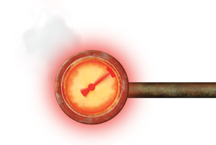
Elevated serum bile acid
- High sBA may be a surrogate biomarker for native liver survival in PFIC 1 and PFIC 2.1,2*
- In ALGS, sBA levels are not always evaluated routinely; however, total bilirubin, ALT, AST, APRI, and CB have been identified as additional prognostic markers for native liver survival.3
- *Based on retrospective analyses of 130 PFIC 1 and 264 PFIC 2 patients from the NAPPED consortium.
Cholestatic pruritus, or itch
Cholestatic pruritus may lead to life-altering symptoms, such as cutaneous mutilation, difficulty sleeping, and impaired school performance.4,5
Failure to thrive
When a child with ChLD has a height and weight that are significantly below that of other children of similar age and sex, they may be diagnosed with failure to thrive.4,6-9
Malabsorption, malnutrition, and FSV deficiency are likely the main causes of these growth impairments in PFIC and ALGS.4,6-9


For illustrative purposes only. Does not reflect a liver with ChLD.
Other indicators of ChLD include jaundice and discolored stools
Jaundice occurs when bilirubin, a biliary pigment, accumulates in tissues like the skin, eyes, and mucus membranes. In ChLD, this happens due to improper elimination of biliary substances in the liver.8,10
Excretory symptoms are also common, including dark urine and pale, discolored, or acholic stools.11
These manifestations lead to an array of physical symptoms and emotional distress, severely impacting quality of life of both patients and caregivers4,7,10,12-15
sBA is one possible indicator of ChLD – and may play a role in pruritus
sBA levels in types of ChLD
Laboratory findings show patients with PFIC and ALGS may present with elevated levels of sBA4,8,9,16


Elevated sBA may be linked to
pruritus, a signature symptom in PFIC and ALGS.8,9,16-18
sBA and pruritus
Hypothesized pathogenesis of pruritus
There are numerous possible pruritogens involved in pruritus, including bile acids, endogenous opioids, and steroid metabolites. While the exact causes of cholestatic pruritus are unclear, one hypothesis is a causative relationship between bile acids and pruritus through multiple pathways.19
Cholestatic Liver
Bile acids in the liver build up, then spill over into the bloodstream12
Skin
Nerve ends under the skin are stimulated19
Brain
Nerves send itch signals to the spinal cord and brain19
Itch
Increased pruritic response19
It is important to know that severity of pruritus differs from patient to patient and may not always directly correlate with their sBA levels.10,17,20
Pruritus is a classic warning sign of ChLD that may warrant serious and prompt attention12,21
ALT=alanine aminotransferase; APRI=aspartate aminotransferase to platelet ratio index; AST=aspartate aminotransferase; CB=conjugated bilirubin; ChLD=cholestatic liver diseases.
References: 1. van Wessel DBE, Thompson RJ, Gonzales E, et al. Impact of genotype, serum bile acids, and surgical biliary diversion on native liver survival in FIC1 deficiency. Hepatology. 2021;74(2):892-906. 2. van Wessel DBE, Thompson RJ, Gonzales E, et al. Genotype correlates with the natural history of severe bile salt export pump deficiency. J Hepatol. 2020;73(1):84-93. 3. Vandriel SM, Li L-T, She H, et al. Natural history of liver disease in a large international cohort of children with Alagille syndrome: results from the GALA study. Hepatology. 2023;77(2):512-529. 4. Srivastava A. Progressive familial intrahepatic cholestasis. J Clin Exp Hepatol. 2014;4(1):25-36. 5. Kamath BM, Abetz-Webb L, Kennedy C, et al. Development of a novel tool to assess the impact of itching in pediatric cholestasis. Patient. 2017;11:69-82. 6. Pawlikowska L, Strautnieks S, Jankowska I, et al. Differences in presentation and progression between severe FIC1 and BSEP deficiencies. J Hepatol. 2010;53(1):170-178. 7. Kamath BM, Baker A, Houwen R, Todorova L, Kerkar N. Systematic review: the epidemiology, natural history, and burden of Alagille syndrome. J Pediatr Gastroenterol Nutr. 2018;67(2):148-156. 8. Krantz ID, Piccoli DA, Spinner NB. Alagille syndrome. J Med Genet. 1997;34(2):152-157. 9. Turnpenny PD, Ellard S. Alagille syndrome: pathogenesis, diagnosis, and management. Eur J Hum Genet. 2012;20(3):251-257. 10. Gunaydin M, Bozkurter Cil AT. Progressive familial intrahepatic cholestasis: diagnosis, management, and treatment. Hepat Med. 2018;10:95-104. 11. Hilscher MB, Kamath PS, Eaton JE. Cholestatic liver diseases: a primer for generalists and subspecialists. Mayo Clin Proc. 2020;95(10):2263-2279. 12. Kamath BM, Stein P, Houwen RHJ, Verkade HJ. Potential of ileal bile acid transporter inhibition as a therapeutic target in Alagille syndrome and progressive familial intrahepatic cholestasis. Liver Int. 2020;40(8):1812-1822. 13. Mighiu C, O’Hara S, Ferri Grazzi E, et al. Impact of progressive familial intrahepatic cholestasis on caregivers: caregiver-reported outcomes from the multinational PICTURE study. Orphanet J Rare Dis. 2022;17(1):1-9. 14. Elisofon SA, Emerick KM, Sinacore JM, Alonso EM. Health status of patients with Alagille syndrome. J Pediatr Gastroenterol Nutr. 2010;51(6):759-765. 15. Quadrado L, Mogul DB, Gurevich A, et al. Caregiver burden associated with caring for a child with Alagille syndrome: a multi-national, quantitative analysis. Presented at: North American Society for Pediatric Gastroenterology, Hepatology and Nutrition (NASPGHAN) Annual Meeting; October 12-15, 2022; Orlando, FL, USA. 16. Davit-Spraul A, Gonzales E, Baussan C, Jacquemin E. Progressive familial intrahepatic cholestasis. Orphanet J Rare Dis. 2009;4:1. 17. Ibrahim SH, Kamath BM, Loomes KM, Karpen SJ. Cholestatic liver diseases of genetic etiology: advances and controversies. Hepatology. 2022;75(6):1627-1646. 18. Mehl A, Bohorquex H, Serrano M-S, Galliano G, Reichman TW. Liver transplantation and the management of progressive familial intrahepatic cholestasis in children. World J Transplant. 2016;6(2):278-290. 19. Imam MH, Gossard AA, Sinakos E, Lindor KD. Pathogenesis and management of pruritus in cholestatic liver disease. J Gastroenterol Hepatol. 2012;27(7):1150-1158. 20. Ayoub MD, Kamath BM. Alagille syndrome: diagnostic challenges and advances in management. Diagnostics (Basel). 2020;10(11):1-18. 21. Tessitore M, Sorrentino E, Schiano Di Cola G, Colucci A, Vajro P, Mandato C. Malnutrition in pediatric chronic cholestatic disease: an up-to-date overview. Nutrients. 2021;13(8):1-23.